MRI Brain Imaging Identifies Regions Responsive to Fatty Foods
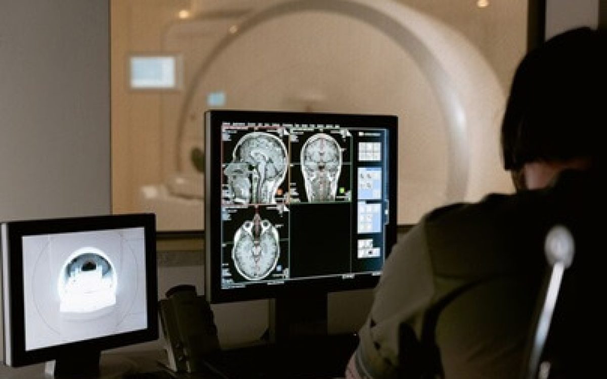
In a study conducted by researchers from the University of Cambridge and the Addenbrooke’s Hospital in the United Kingdom, the team successfully identified regions in the brain that respond to the consumption of foods high in fat content using magnetic resonance imaging (MRI).
As part of the study, volunteers were subjected to MRI scans while consuming doses of whipped cream with varying fat percentages. The research team recruited 22 volunteers who participated in the experiment and consumed different doses of whipped cream, with MRI scans of their brains conducted during the course of the experiment.
The researchers found that the anterior cingulate cortex, located within the brain, lit up on the MRI scans when participants consumed fatty foods during the experiment. The intensity of illumination in this brain region on the MRI images increased with the consumption of higher-fat content beverages.
It is worth noting that previous studies have demonstrated that excessive consumption of high-fat foods is a key contributing factor to obesity.
The researchers aim to pinpoint the areas within the brain that respond to fat content, enabling the development of therapeutic methods to reduce the natural inclination to consume such high-fat foods.


